Scanning Name
CT ABDOMEN AND PELVIS
Reports
Fast report turn around time
Laboratory Professionals
Specialised clinical reports by experts
CT Abdomen and Pelvis Imaging
A CT scan of the abdomen and pelvis is a comprehensive imaging procedure that visualizes the organs and structures in the abdominal and pelvic regions. It helps in diagnosing a wide range of conditions affecting the digestive, urinary, and reproductive systems.
Process:
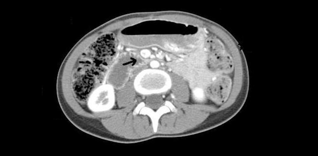
During a CT abdomen and pelvis scan, the patient lies on a table that moves into the CT scanner. The scanner captures detailed cross-sectional images of the abdomen and pelvis using X-ray technology. These images are processed by a computer to create detailed pictures of abdominal organs, blood vessels, lymph nodes, and pelvic structures.
Uses:
- Diagnosis of Abdominal Pain: CT imaging assists in diagnosing the cause of abdominal pain, such as appendicitis, pancreatitis, or gastrointestinal perforation.
- Evaluation of Organ Health: It helps in assessing the liver, spleen, pancreas, kidneys, and adrenal glands for tumors, cysts, or other abnormalities.
- Detection of Abdominal Masses: CT scans can detect and characterize masses or tumors in the abdomen or pelvis.
- Assessment of Trauma: CT imaging is crucial for evaluating abdominal injuries following accidents or trauma.
- Staging of Cancer: CT scans aid in cancer staging by determining the extent of tumor spread within the abdomen or pelvis.
Others:
- Comprehensive Diagnostic Tool: CT abdomen and pelvis provide comprehensive views of multiple organ systems, aiding in accurate diagnosis and treatment planning.
- Visualization of Blood Flow: CT angiography can be performed to visualize blood vessels and detect abnormalities like aneurysms or arterial stenosis.
- Guidance for Surgical Planning: Surgeons use CT images for planning abdominal or pelvic surgeries, such as tumor resections or organ transplants.
